ePoster

QR unavailable
Share ePoster
Scan or copy the public World Wide URL.
ePoster
ASTROGLIAL ORGANIZATION IN THE NEUROVASCULAR UNIT IN HUMAN POSTMORTEM BRAIN TISSUE: A QUANTITATIVE ANALYSIS BY CONFOCAL MICROSCOPY
América Vera-Montecinosand 4 co-authors
Psiquiatria Molecular MERITT, Institut de Recerca Sant Joan de Déu, Barcelona, Spain; Departamento de Ciencias Biológicas y Químicas, Facultad De Ciencias, Universidad San Sebastián, Concepción, Chile
FENS Forum 2026 (2026)
Barcelona, Spain
Presenter and authors
Presenter
América Vera-Montecinos
Psiquiatria Molecular MERITT, Institut de Recerca Sant Joan de Déu, Barcelona, Spain; Departamento de Ciencias Biológicas y Químicas, Facultad De Ciencias, Universidad San Sebastián, Concepción, Chile
Co-authors
Mónica Roldán; Marcos Frias; Ioannis Georvasilis; Belén Ramos
Abstract
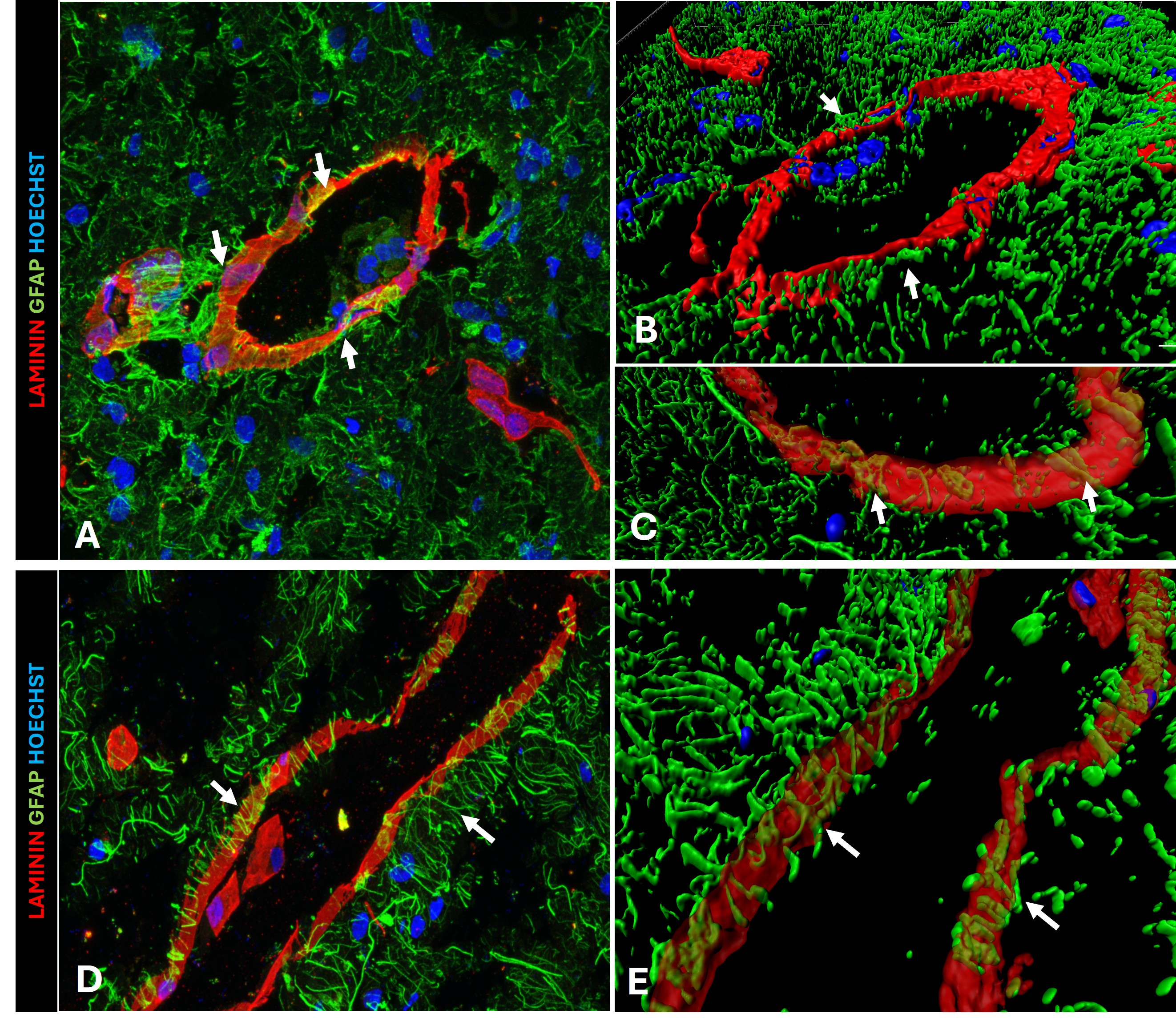
Astroglia projects end-feet to vascular basal membrane at the neurovascular unit. However, limited information is available on how is organized the glial projections in human brain and quantification approaches to measure the degree of organization has not been developed yet. The aim of this study was to develop a method for quantify astroglial organization in human cerebellar cortex. Postmortem human cerebellar frozen tissue (n=5) was cut in a cryostat to obtain 7 μm thick sagittal sections. Immunofluorescence labelling with anti-laminin and anti-GFAP was used. Confocal and super-resolution microscopy analysis was performed by Leica TCS SP8 equipped with a white light laser and Hybrid spectral detectors. 3D reconstructions were generated. For colocalization analysis the JACoP plugin in ImageJ (NIH), version 1.53q, was used. Manders’ coefficients (M1 and M2) and the intensity correlation quotient (ICQ) were calculated. Co-labelling with GFAP and laminin detected perpendicular well-organized glial projections with numerous contacts with basal lamina in all tissue samples. 3D reconstructions revealed multiple end-feet contacts surrounding blood vessels. Quantitative colocalization analysis found a high overlapping between GFAP with vessels and a low variation across different brain tissue samples of independent subjects. This study provides the foundations for the visualization of the astroglia organization in the neurovascular unit providing the first attempt to quantify the organization of astroglia at the neurovascular unit. Thus, this advanced methodology will be applied in different pathologies to measure the degree of organization of these structures. CP16/00153 and PI21/00059 to BR (Instituto de Salud Carlos III.