ePoster

QR unavailable
Share ePoster
Scan or copy the public World Wide URL.
ePoster
SURGICAL ACCESS AND ANATOMICAL–HISTOLOGICAL CHARACTERIZATION OF THE PUDENDAL NERVE IN PIGS
Naif Onur Ceylanand 11 co-authors
University of Pisa
FENS Forum 2026 (2026)
Barcelona, Spain
Presenter and authors
Presenter
Naif Onur Ceylan
University of Pisa
Co-authors
Filippo Castellani; Alice Giannotti; Giulia Lazzarini; Khatia Gabisonia; Chiara Di Franco; Irene Nocera; Andrea Pirone; Alberto Elmi; Angela Briganti; Silvestro Micera; Vincenzo Miragliotta
Abstract
Lower urinary tract (LUT) disorders significantly impair quality of life and social well-being. To address these conditions, various surgical and bioengineering strategies have been developed using the pig as an experimental model due to its close anatomical and dimensional similarity to humans.
The pudendal nerve contains both somatic and autonomic fibers and plays a central role in pelvic motor and sensory function. A detailed anatomical and histological characterization is therefore critical to enable targeted neuromodulation strategies for the management of lower urinary tract (LUT) dysfunction.
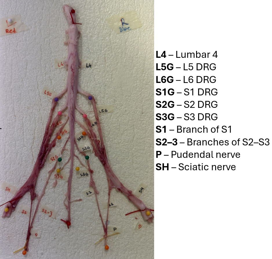
In this study, we characterized surgical access to the pudendal nerve, its root-level topography, and its microstructural organization in mini pigs (n = 4) and farm pigs (n = 15). We first assessed a caudolateral intermuscular approach for pudendal nerve exposure. Following euthanasia, the pelvic region was dissected, and the nerve was traced from the sacral plexus to its terminal branches. Tissue samples were collected for histology and stained with hematoxylin–eosin, Luxol Fast Blue, and Goldner’s Trichrome.
The transgluteal approach enabled reliable access to the pudendal nerve with minimal tissue disruption, achieved through blunt dissection between the musculus gluteus superficialis, musculus biceps femoris, and musculus gluteus medius. Histological analysis revealed that the pudendal nerve contains ~50 fascicles, and distinct root-level branching patterns were observed between pig types. Together, these results provide a detailed morphological and topographic map of the porcine pudendal nerve, establishing a robust anatomical basis for future bioengineering and neuromodulation studies.
The pudendal nerve contains both somatic and autonomic fibers and plays a central role in pelvic motor and sensory function. A detailed anatomical and histological characterization is therefore critical to enable targeted neuromodulation strategies for the management of lower urinary tract (LUT) dysfunction.
In this study, we characterized surgical access to the pudendal nerve, its root-level topography, and its microstructural organization in mini pigs (n = 4) and farm pigs (n = 15). We first assessed a caudolateral intermuscular approach for pudendal nerve exposure. Following euthanasia, the pelvic region was dissected, and the nerve was traced from the sacral plexus to its terminal branches. Tissue samples were collected for histology and stained with hematoxylin–eosin, Luxol Fast Blue, and Goldner’s Trichrome.
The transgluteal approach enabled reliable access to the pudendal nerve with minimal tissue disruption, achieved through blunt dissection between the musculus gluteus superficialis, musculus biceps femoris, and musculus gluteus medius. Histological analysis revealed that the pudendal nerve contains ~50 fascicles, and distinct root-level branching patterns were observed between pig types. Together, these results provide a detailed morphological and topographic map of the porcine pudendal nerve, establishing a robust anatomical basis for future bioengineering and neuromodulation studies.